Software Vendors & OEMs
Ready to integrate web DICOM PACS Viewer and connectivity solutions

HTML 5
ZERO-FOOTPRINT
DICOM VIEWER
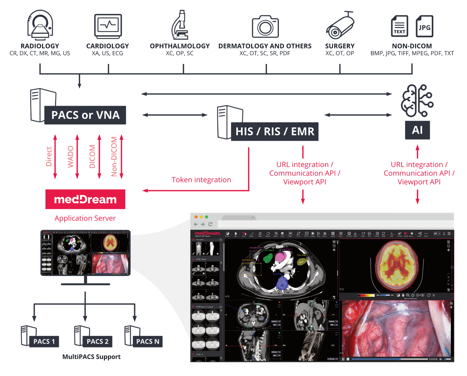
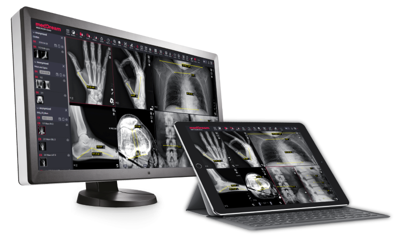
MedDream is a web-based DICOM Viewer for PACS server that is aimed at making diagnoses, viewing, archiving and transmitting medical images. The DICOM Viewer can be integrated into any PACS system/VNA including: MedDream PACS, PacsOne, dcm4chee, Orthanc, Conquest, ClearCanvas, Google Cloud Healthcare, etc.
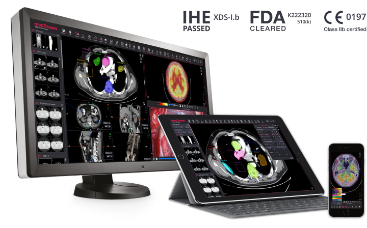
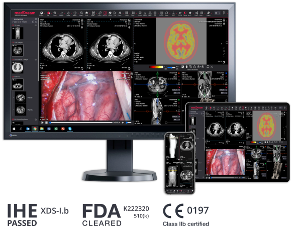
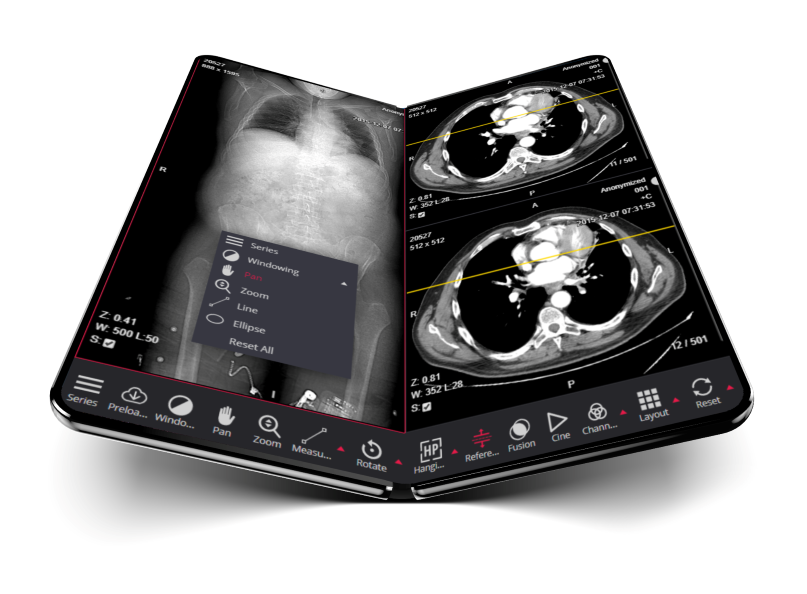
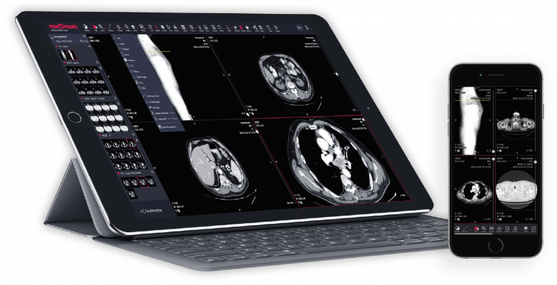


The MedDream DICOM Viewer ensures a prompt and reliable way to search, view, analyze and diagnose medical images, signals and video files from anywhere and on any device. Web-based DICOM Viewer is developed using responsive design, that enables medical images to be accessed not only on computers, but also on tablets, smart phones or other devices with mobile view functionality.
MedDream is FDA cleared for diagnostic use including mammographic images and is CE Class IIb certified as a medical device that can be used for review purposes or even primary diagnosis. Read more about MedDream certifications.
As a diagnostic image viewer, MedDream consists of a Viewer component, which runs in a browser and does not require any installation on the client device, and a MedDream application server, which handles communication with the hospital systems (HIS/RIS/PACS/VNA and any other EMR) and prepares images for streaming to the MedDream DICOM Viewer.
MedDream uses a flexible and open integration interface for connecting to HIS and/or EMR systems primarily based on URL calls, thus allowing it to be integrated into any medical application.
Integration of the web DICOM Viewer:
URL link examples for DICOM Viewer integration into Information System:
Integration options:
Read more about MedDream DICOM Viewer integration into web applications in Integration Guide.
The MedDream DICOM Viewer supports the following DICOM files and formats: Implicit Little Endian, Explicit Little Endian, JPEG, JPEG-LS, JPEG 2000, RLE, MPEG-2 and MPEG-4 Transfer Syntaxes.
The html5 zero-footprint DICOM Viewer, MedDream, can be integrated into any PACS system/VNA including: MedDream PACS, PacsOne PACS Server, Orthanc PACS, open-source: dcm4chee PACS, Conquest DICOM Software, ClearCanvas, etc. MedDream also can be integrated into cloud DICOM services: Google Cloud Healthcare, Azure Health Data Services, Amazon AWS S3 storage, etc.
Available versions of the DICOM Viewer for PACS servers:
The MedDream DICOM Viewer also supports MultiPACS by PACS plugins: dcm4chee v2, dcm4chee v5, Orthanc, PacsOne, DICOM QueryRetrieve, FileSystem.
The MedDream application server connectivity to the PACS can be achieved using:
MedDream supports Cross-enterprise Document Sharing for Imaging (XDS-I.b) profile as DOCUMENT CONSUMER (ITI-18, ITI-43, RAD-16, RAD-55, RAD-69 transactions). Read more about MedDream DICOM Viewer integration with IHE XDS profiles.
Depending on each specific installation, the preliminary hardware sizing information may vary slightly based on the modalities used in the institution. The minimum hardware requirements for the MedDream Application Server are described in Minimal server side requirements page.
The MedDream, html5 zero-footprint DICOM Viewer for PACS server/VNA, ensures a prompt and reliable way to search, view, analyze and diagnose medical images, signals and video files from anywhere and on any device: computers, tablets and smart phones.
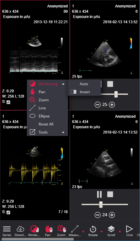
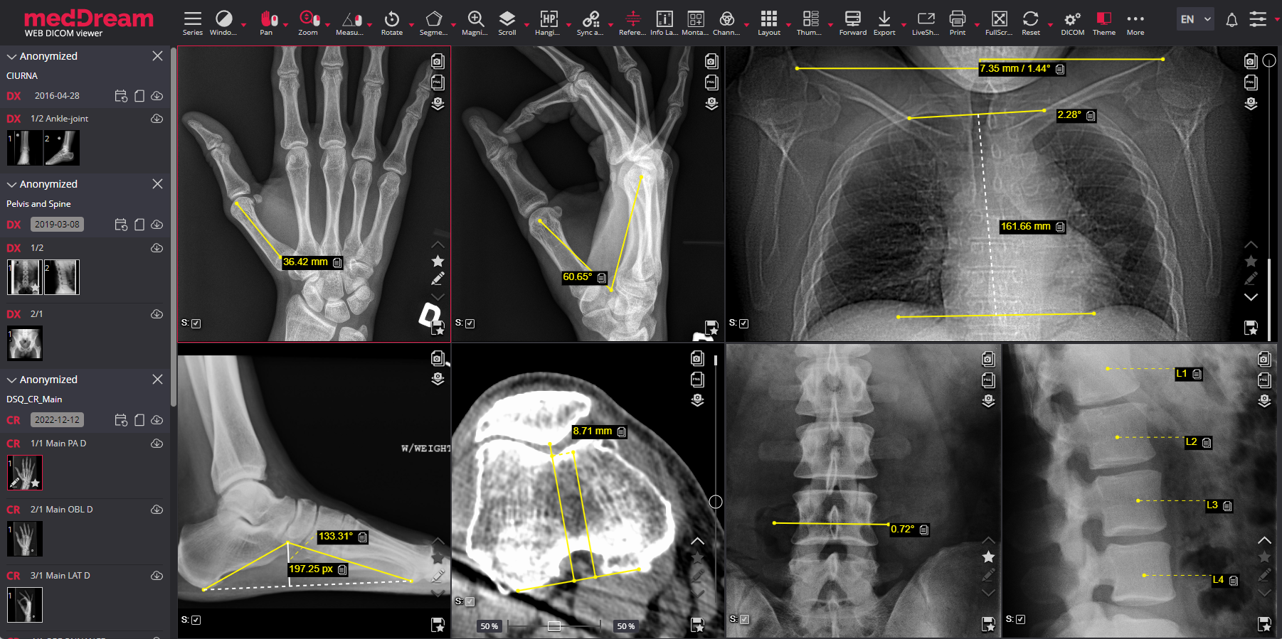
The MedDream DICOM Viewer has an extensive radiology tool set, which includes regular features such as:

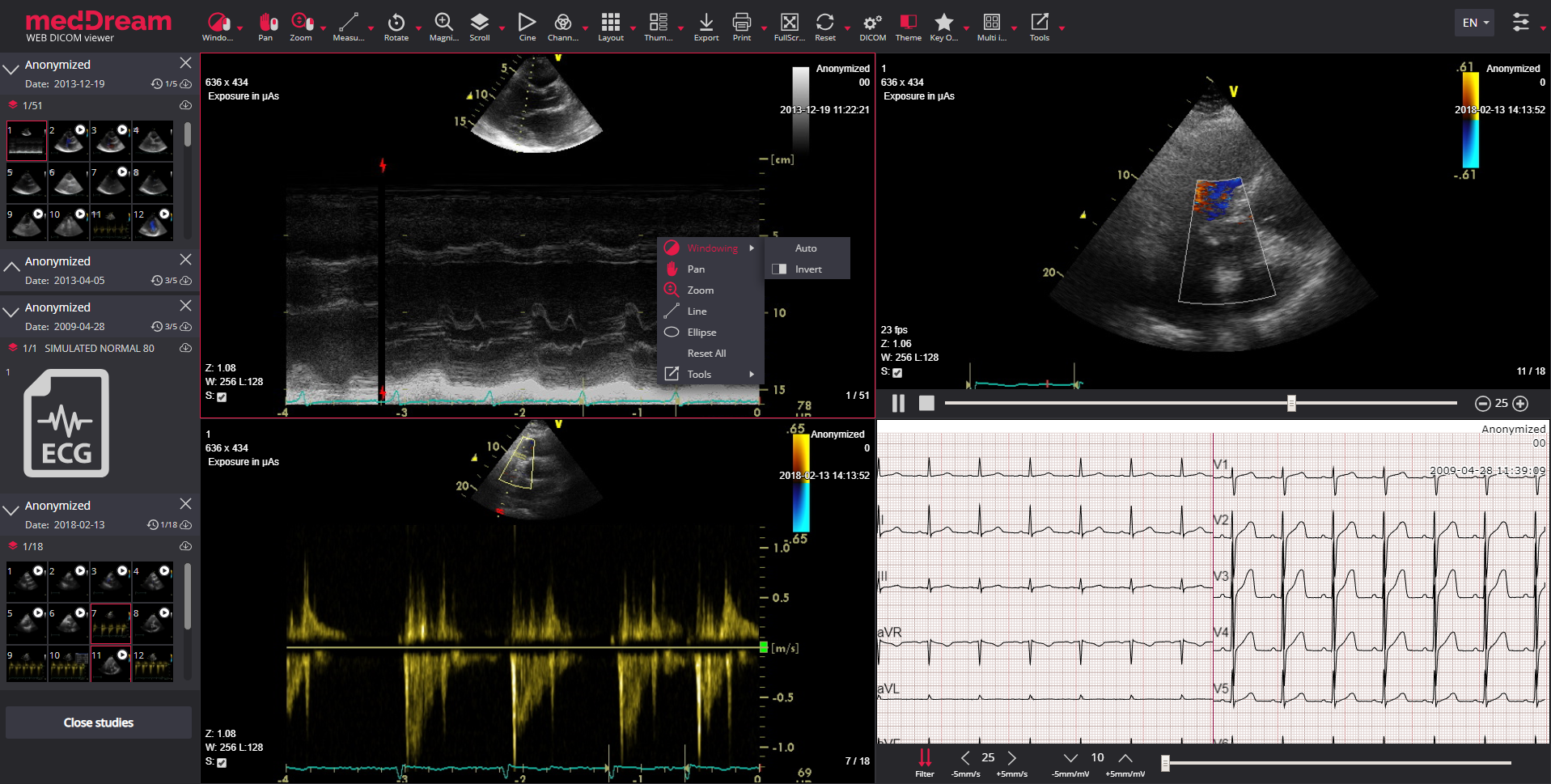
Window. Image window level manipulation using the mouse or selecting from a list of available presets;

Pan. Moving the image within the image viewer, useful when image is larger than the viewer, after zooming for example;

Zoom. Increase/decrease the image;

Scroll. Scroll through the images of series by using mouse wheel, dragging vertically or with keyboard hot keys;

Rotate/Flip. Rotate the image right/left, flip it horizontal/vertical with possibility to revert the image to original orientation;

Magnifier. Magnify (enlarge) certain area of the image.
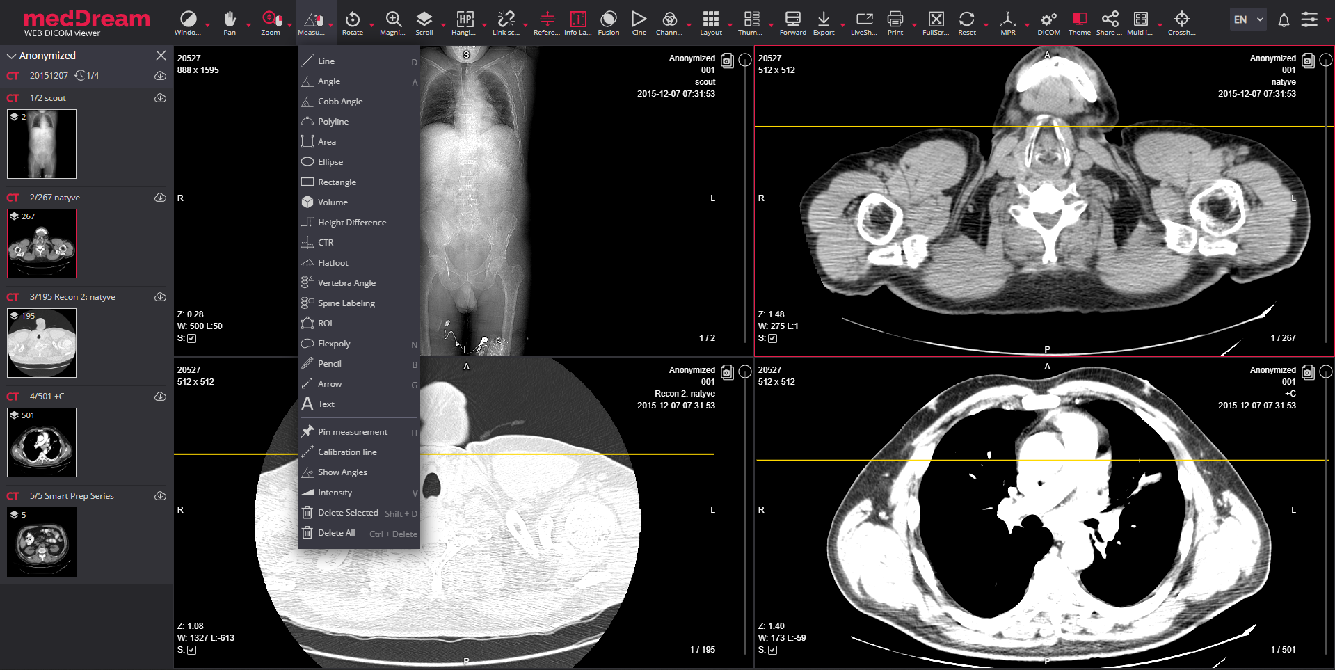
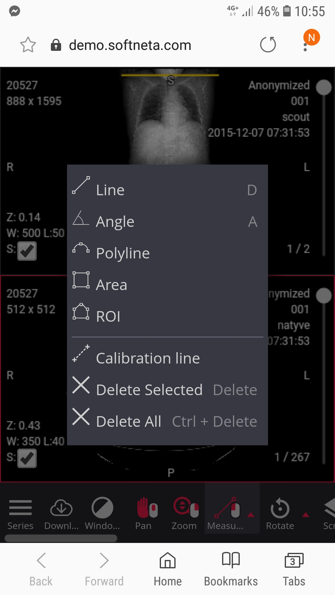
The MedDream DICOM Viewer advanced features and radiology measurements are:

Line. Draw and measure the length of a line;

Angle. Draw and measure an angle;

Cobb angle. Draw and measure a Cobb angle;

Polyline. Draw and measure the length of a polyline;

Area. Mark area of interest with a polyline and measure its area;

Ellipse. Measure an ellipse area, min, max, mean, and standard deviation values;

Rectangle. Measure a rectangle area, min, max, mean, and standard deviation values;

Volume. Measure the volume of a 2D image. The 2D area is spun around a selected axis to form a 3D shape and the volume of the shape is then measured;

Height Difference. Measure the vertical distance and angle between the horizontal line and the line connecting the points;

CTR. Measure a cardiothoracic ratio to estimate a heart size;

Flatfoot. Foot longitudinal arch measurement to detect longitudinal flatfoot;
Goniometry. Measure the lengths of the femur, tibia, and femoral-tibial angular deformities;
TT-TG distance. Measure the tibial tuberosity to trochlear groove distance for quantifying the knee patellar instability;

Spine labeling. Measurement to mark the vertebrae of the spine;

Vertebra Angle. Measure an angle between the user-drawn vertebra axis and the horizontal axis of the image;

Time-Intensity curve. Visualize the lesions' behavior by plotting the ROI intensity values over time after the administration of contrast material;

ROI. Mark and store ROI for study instance;

Closed Polygon. The ROI with curved lines;

Flexpoly. Mark flexible region of the image for which the area is calculated;

Pencil. Mark the area of the image with a free drawing;
Arrow. Mark the area of interest on image, video or multi-frame;

Text. Possibility to write, view, edit or delete text;

Continuous measurement. Possibility to activate the tool in order to repeat measurements several times.

Repulsor. The tool to adjust Closed Polygon;

Intensity. Measure Hounsfield units at a specific point of a CT study;

Show angles. Show all angles between intersecting lines;

Ruler. Show the rulers at the bottom and on the left side of the viewport;

Calibration line. Change the scale of measurement;

Delete annotation. Possibility to delete all or selected annotations;

Save annotation. Possibility to save and display annotations.

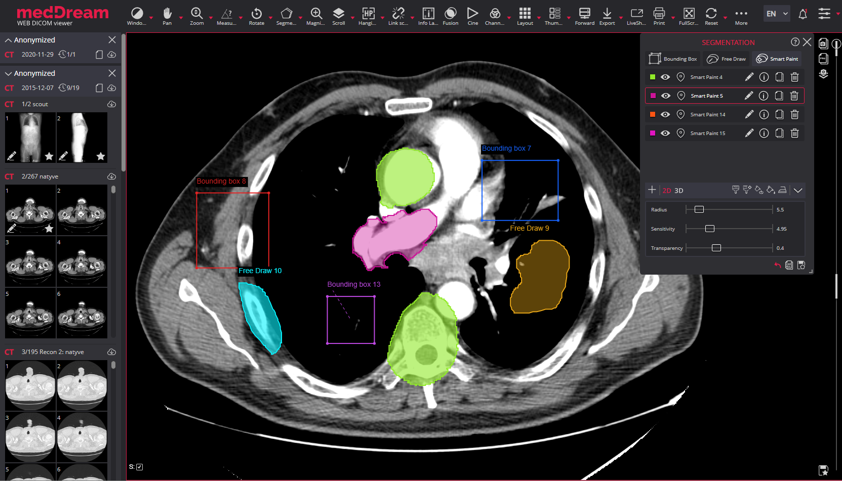
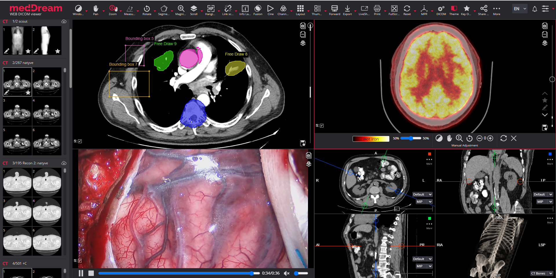
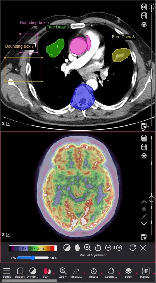
The MedDream DICOM Viewer has Segmentation tools for marking the regions of interest in medical images, saving them:

Bounding Box. The tool allows marking the region of interest by drawing the bounding rectangular around it. Possibility to mark 2D or 3D bounding box segments.

Smart Paint. Marking the region of interest by drawing the contour on the image with free drawing tools. Adjustable radius and sensitivity. Possibility to use 2D and 3D modes.

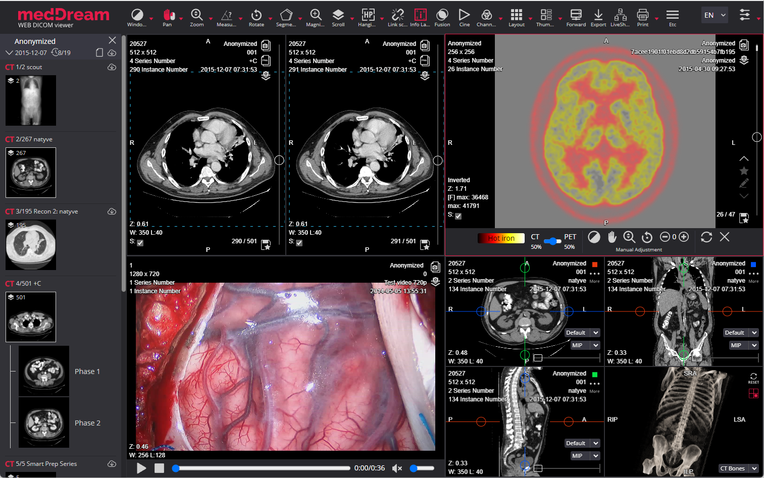
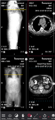
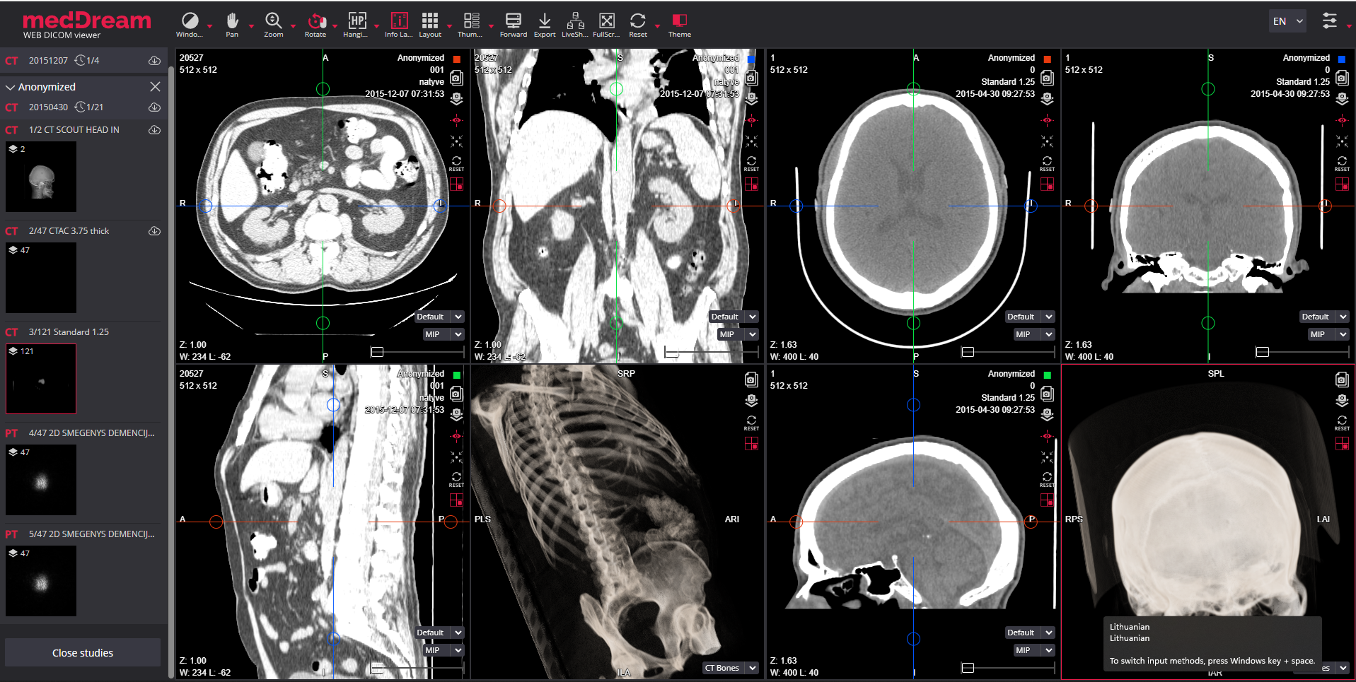
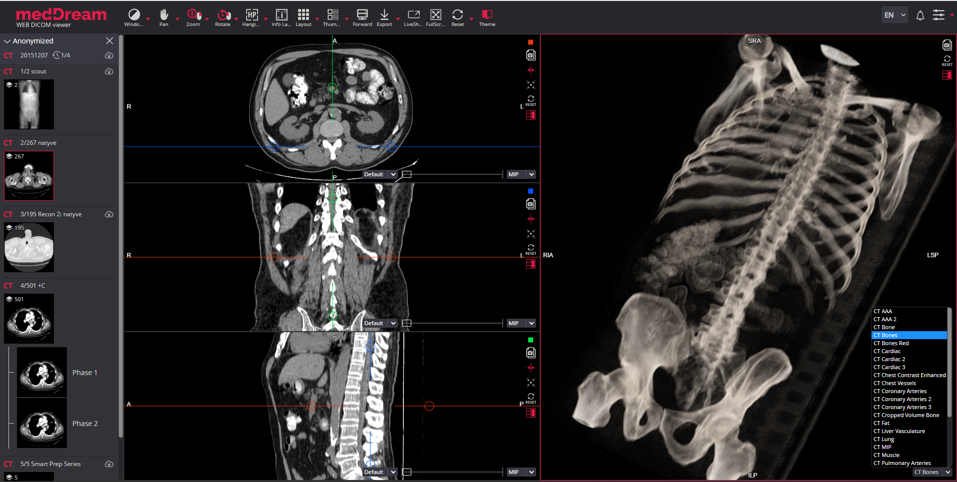
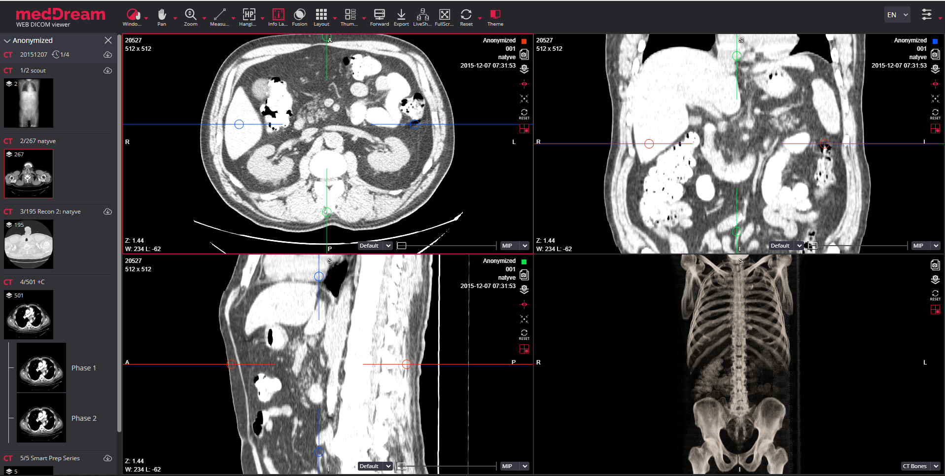
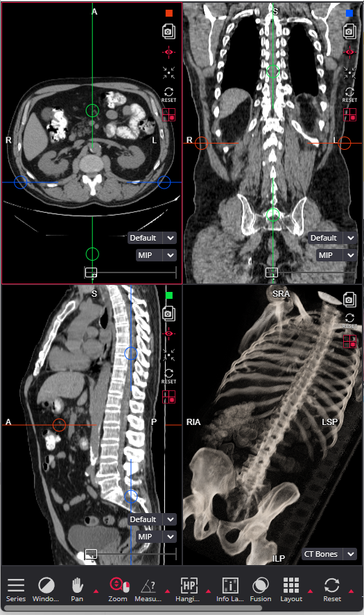
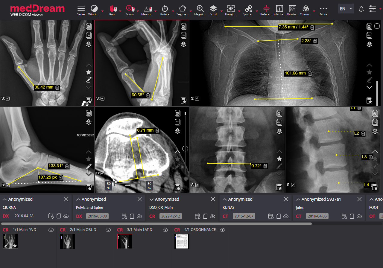
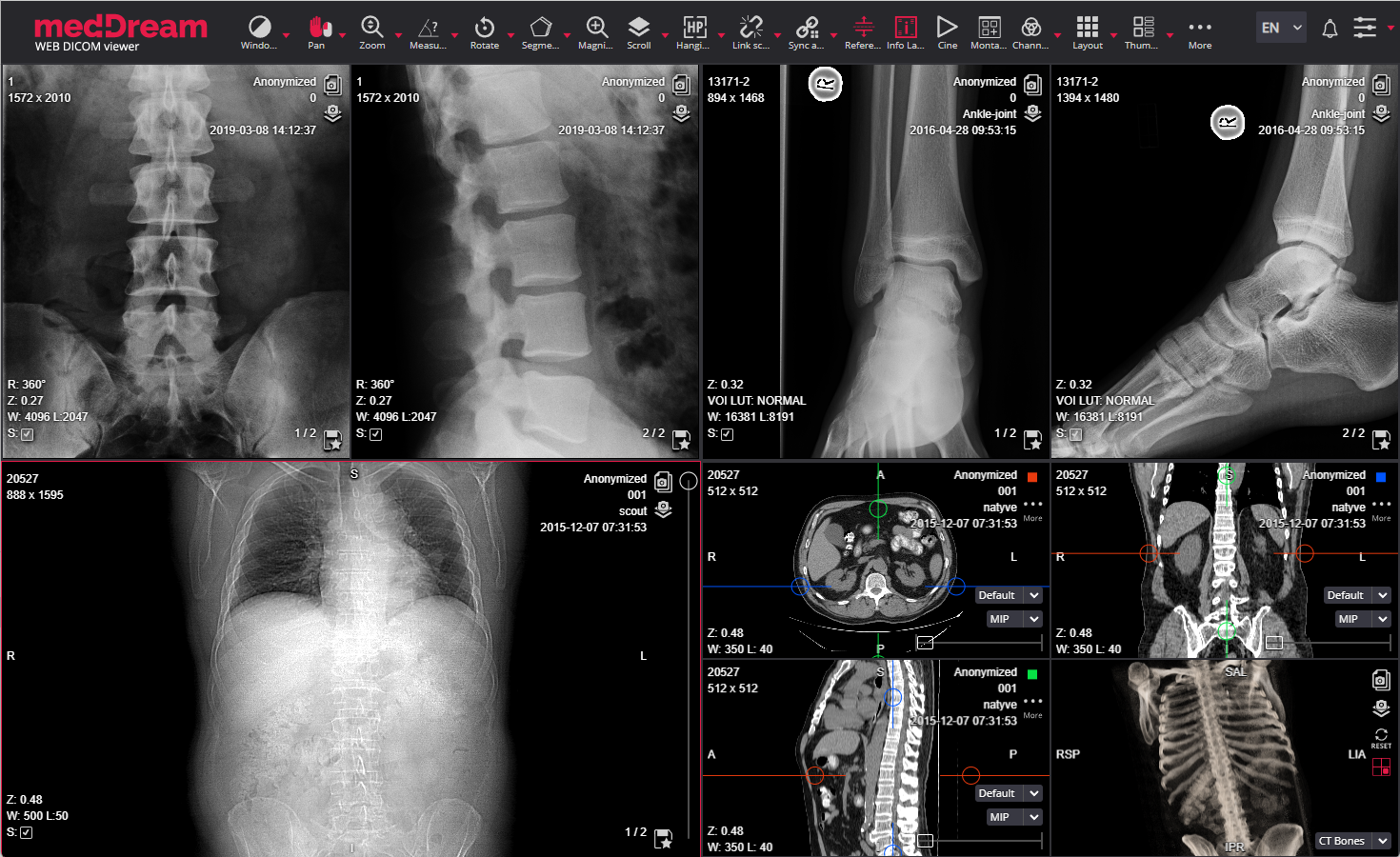
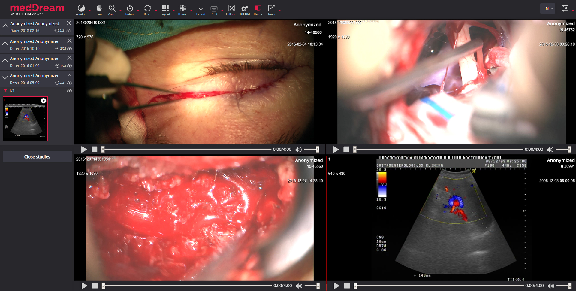
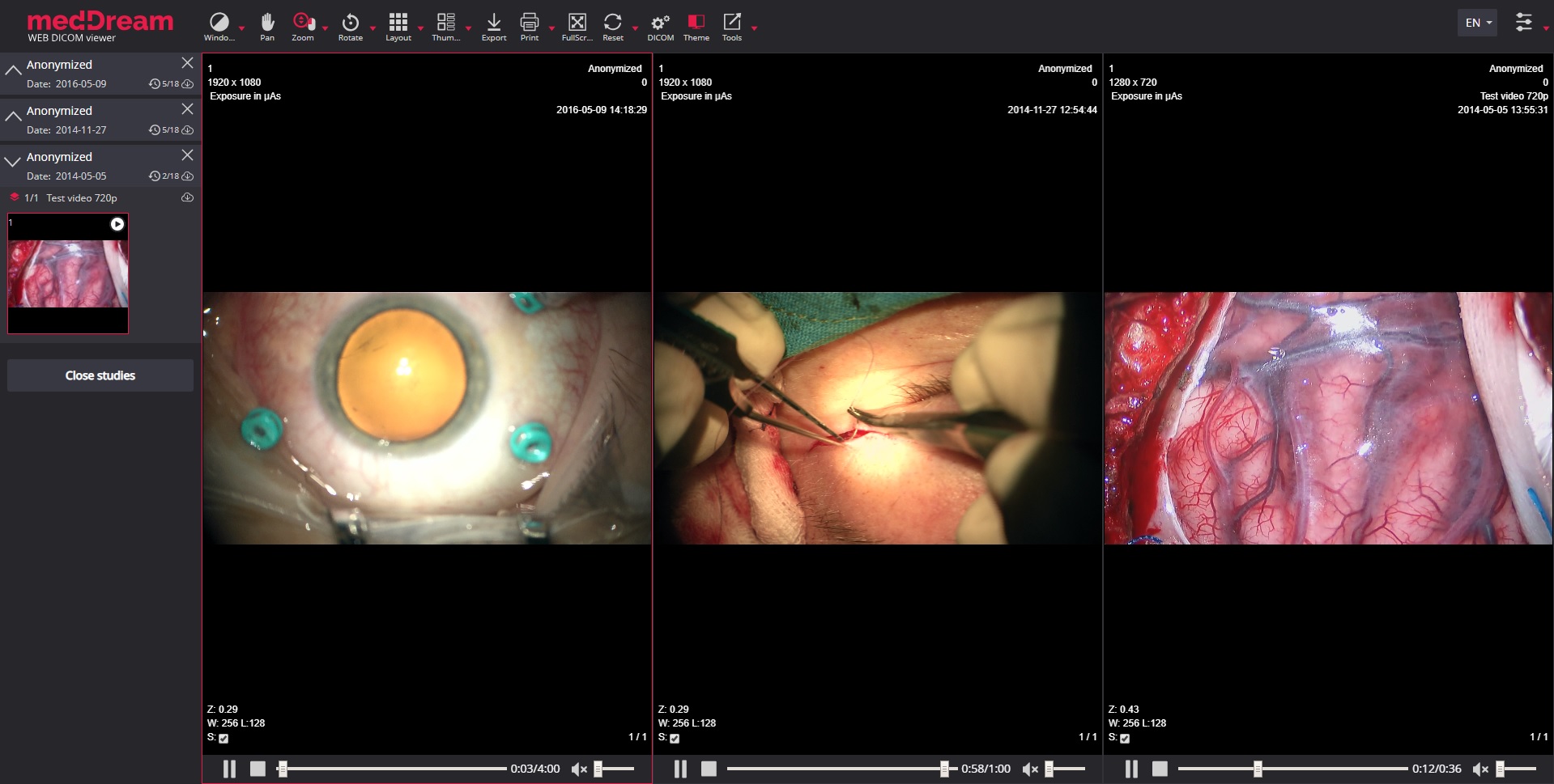
Layout. Select from different types of layouts to view up to 16 DICOM instances at the same time;

Thumbnails. Change position of thumbnails on the screen;

Full Screen. Possibility to switch to full screen view and switch back;

Multi-image/series. Select how many images/series can be loaded in the active viewport;

Split into 2 Panels. Possibility to split viewports into 2 panels;

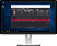
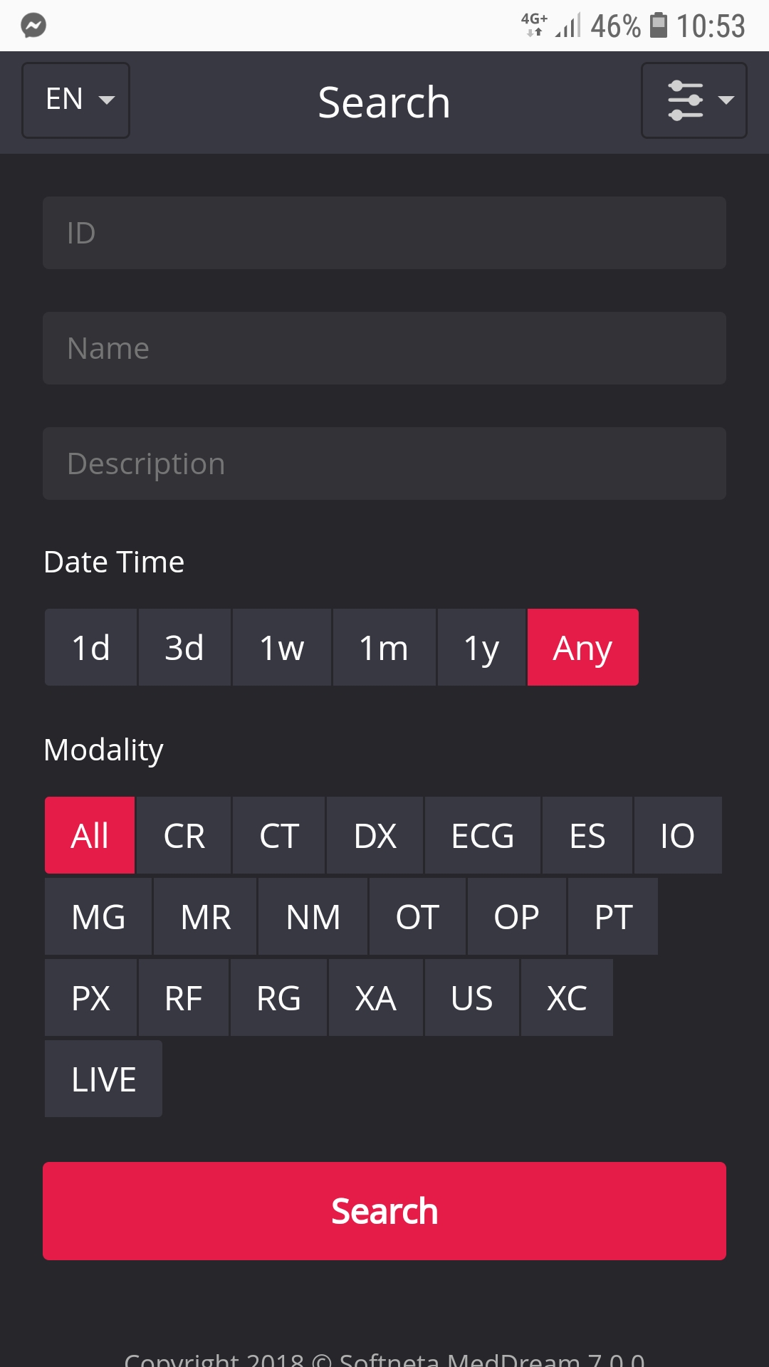
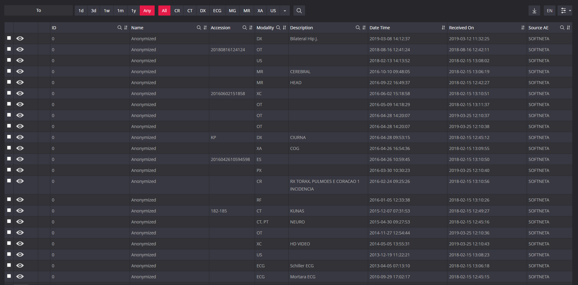
Patient History. Easy access to the entire Patient History. Possibility to filter the studies by ID, Name, Modality, Description, etc. Unique Year filter to filter the studies by the year.

Key Objects storing. Possibility to mark instances as KO and save them for later review;

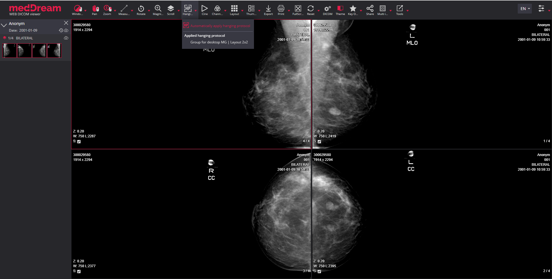
Hanging Protocols. Describe how to lay out a set of DICOM images for faster diagnosis;

Previous Hanging Protocol. Button in the toolbar to quickly apply previous Hanging Protocol.

Next Hanging Protocol. Button in the toolbar to quickly apply next Hanging Protocol.
The DICOM Viewer has also: split-view mode, related studies, multiple studies and multi-monitor support functions. To find more information, please read the product Specification file.
The MedDream DICOM Viewer also has the image manipulation features:

Reference lines. Overlaying reference lines allow to indicate the location of an image slice on another image of an intersecting pane;

Crosshair. Represents the intersecting planes of the selected point on the main study;

Align & Lock. The left or right image alignment and locking function while applying zoom or pan actions. Possibility to move labels and buttons to the opposite side;

Link scrolled series. The possibility to enable/disable simultaneous scrolling for each viewport. The modes of linking the series: Disabled, Manual, Automatic and Distance mode;

Sync Windowing. Optional same-series windowing synchronization;
Sync actions. Sync Windowing, Color Palette, Pan, and Zoom actions for the same series or for all viewports;

Color palette. Possibility to apply color palette for monochrome DICOM images;
Histogram. Showing how the data is distributed across different values for visual Windowing (W/L) changes. Y-axis zooming;

Cine Mode. Cine playback of multi-frame sequences with video seeking support;

Reset. Resetting the image’s view to the original state.
MedDream provides the possibility to select and apply VOI LUT: non-linear transformation created by medical modality.
Rebranding as white label product with vendor’s branding possibility:
Usage simplification:
The main supporting functions of the DICOM Viewer are as follows:

Orientation labels. Labels for tracking of image orientation when manipulating the study with the transformation tools;

Info Labels. Possibility to show/hide Info Labels in viewports;
Report. Possibility to write a report for a study;

Print. Possibility to print an active image;

Forward. Forward several studies at once and easily find devices while forwarding;

Export. Export multiple studies, save in different formats or burn them to CD/DVD/Dual Layer DVD;

Share. Possibility to anonymize and share studies (via DICOM Library).
More supporting functions of the DICOM Viewer, as: search engine, DICOM print, burn, keyboard shortcuts are described in the product Specification file.
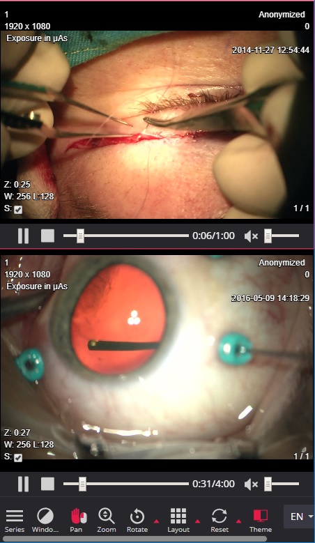
The MedDream DICOM Viewer also has specific features including multi-frame and video support:

Live Share support. Diagnostic quality real-time-sharing functionality (conference mode);

Presenter tool. For marking an area of interest in the viewport during presentation. Presenter tools: Fading line, Fading arrow, Draw, Clear All, Laser Pointer and selecting from 5 colors;

Multi-frame support. The US and XA multi-frames are shown in cine mode. CT/MR/MG/OPT/NM studies multi-frames shown as instance series;

Video support. MPEG-2 and MPEG-4 video support;

ECG support. Electrocardiography study support;

PDF support. Support for PDF modality;

SR support. Support for SR modality;

Non-DICOM support. BMP, JPG, TIFF, MPEG, PDF, and TXT files support;

PR Support. Support for Presentation State annotations;
Key Objects (KO) support. Possibility to mark instances as KO and save them;
Montage. Possibility to create a Montage of different instances and save it as secondary capture in a new series;

PET-CT Fusion. Possibility to combine the series of PET and CT types, thus linking the sites of radioactive drug concentrations with the anatomical patient structure;
Fusion. In addition to the PET series, CT, MR, and NM series may also be used for the Fusion feature;

Construct 4D series. 4D Tool to create virtual series from the study series, where the data are sorted in space and then in time;

Digital Subtraction. Apply Digital Subtraction Angiography mask for XA images;

Color channels. Highlight a color component or a combination of them;

MPR. Multi-planar reconstruction with auto rotate Coronal or Sagittal projections.
The DICOM Viewer’s MPR (multi-planar reconstruction) features are:
MIST MIP features:
All features of the DICOM Viewer and other specifications are described in the product Specification file. For more information, download the MedDream DICOM Viewer brochure.
The MedDream DICOM Viewer’s MPR/MIP/3D functionality simplifies reconstruction technique for three-dimensional visual representations of two-dimensional image slices. The technology offers many different alternate views of the original data using various 3D reconstruction techniques such as MPR and MIP.
MPR Oblique MIST feature with MPR/MIP/3D rendering has the standard tools and advanced MPR/MIP/3D features.
MPR Oblique MIST (client-side rendering) features:

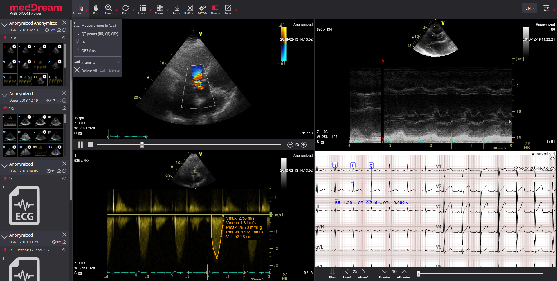
The MedDream DICOM Viewer provides not only standard image manipulation tools but also a way to read, manipulate and interpret electrocardiography (ECG) and ultrasound (US) data.
ECG manipulation tools are all presented in an innovative zoom model, which allows zooming in/out, measure and quantify the ECG data:

Measurement (mV, s). Area calculation indicating beats per minute, time, millivolt (mV, s, bpm);

QT points (RR, QT, QTc). QT interval - the RR interval is calculated as well as QT and the QTc (based on Bazett’s formula);

HR. Measure heart rate (HR) and compare its interval variance over the ECG;

QRS axis. Measure the QRS electrical heart axis;

Studies comparison. Compare of two or more ECGs.
For ultrasound data MedDream also supports these features:

VTI. Velocity Time Integral used to measure the distance which the blood was ejected over a date interval of time;
Up to 12 US studies may be opened at once.
Supported ECG devices:
Other parties’ brand names, with ownerships belonging to Mortara Instrument, custo med GmbH, SCHILLER AG, GE Healthcare, are not authorized by, sponsored by or associated with the MedDream trademark owner.
MedDream can be used to measure the volume of a 2D image by using Simpson’s approximation rule; the 2D area spun around a selected axis to form a 3D shape and the volume is measured. This technique allows volume measurements of a heart in a 2D computed radiography image.
MedDream supports measurement of the Velocity Time Integral (VTI) on ultrasound (US) studies that can quantify the trace of the Doppler flow profile.
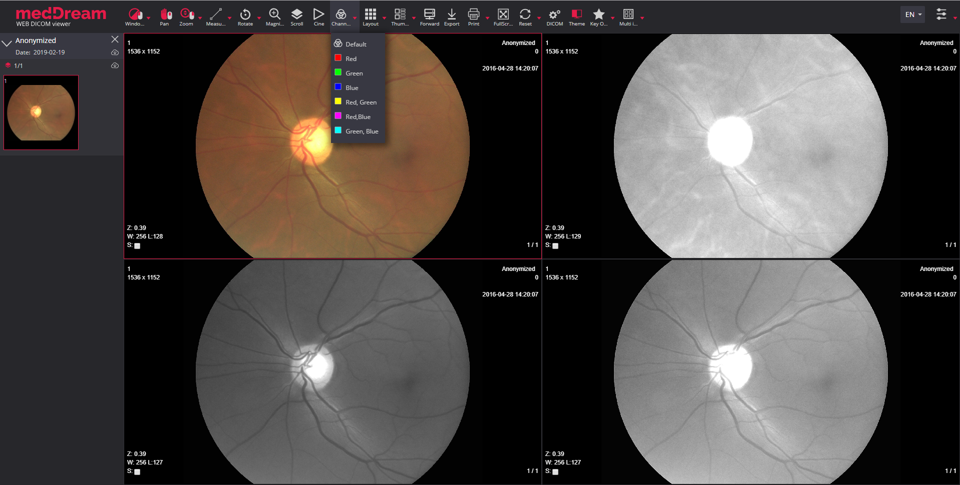
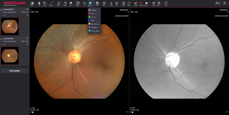
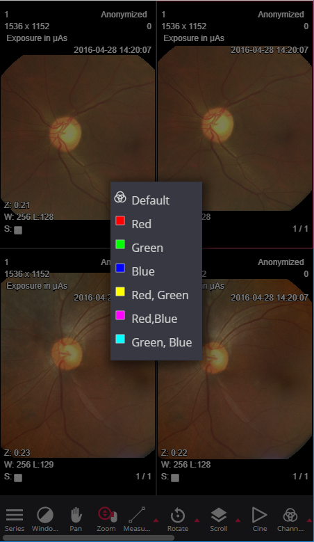
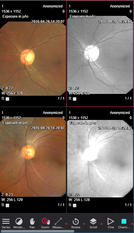
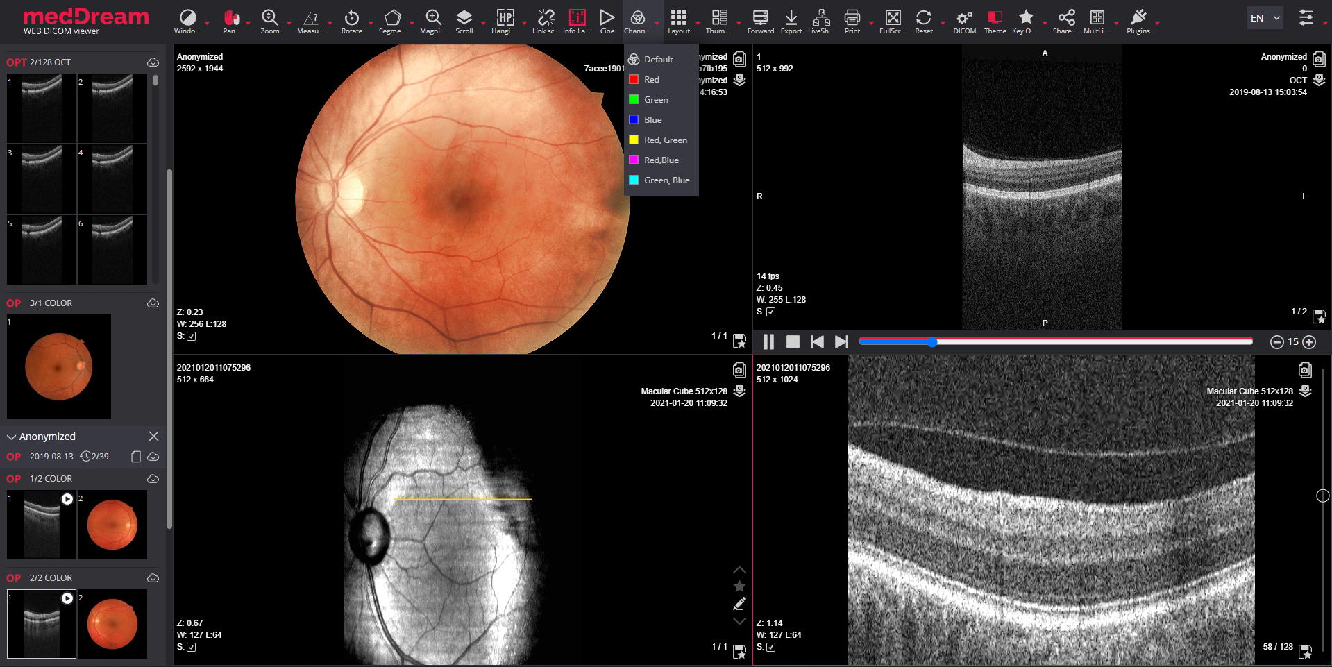
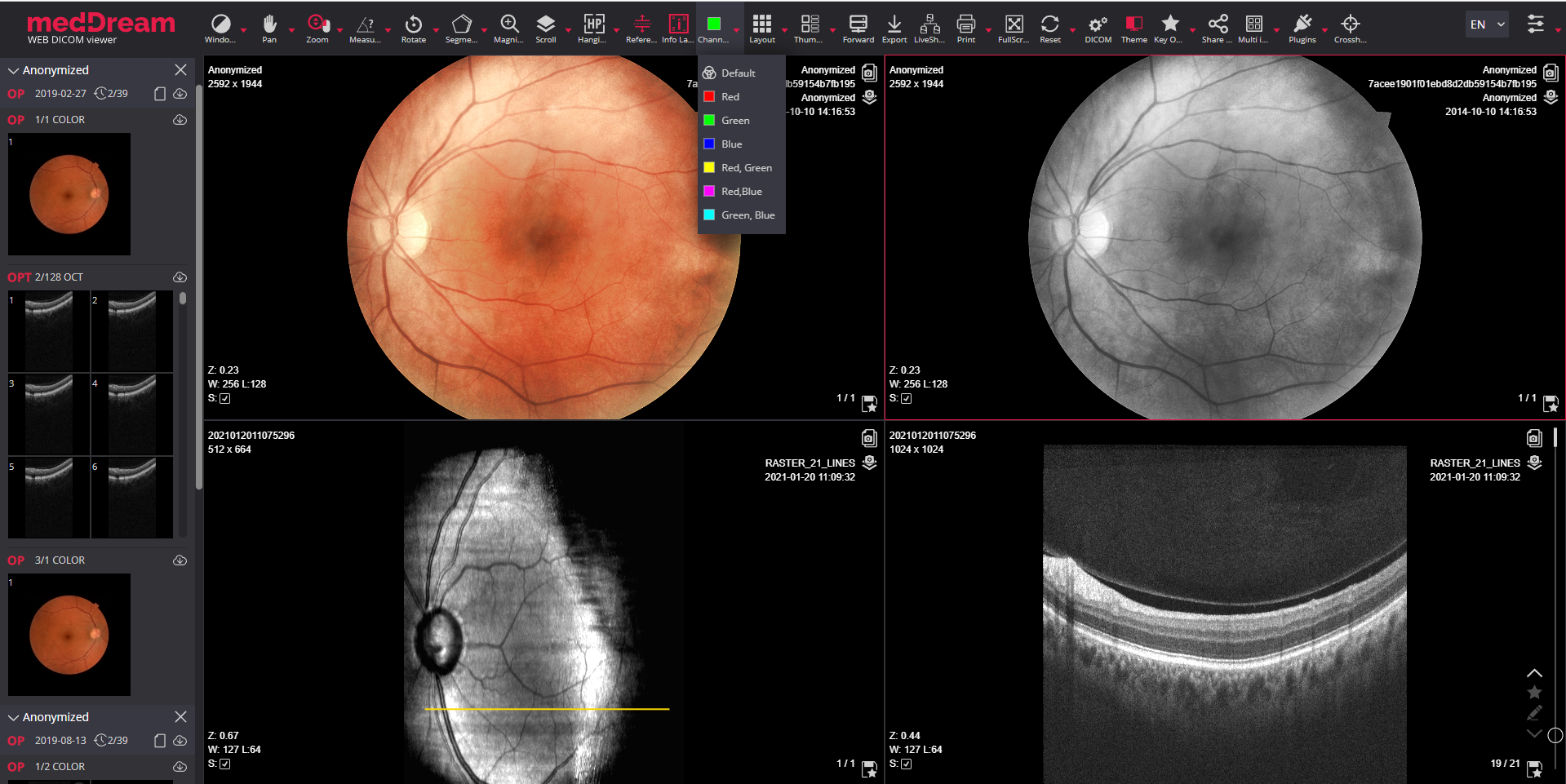
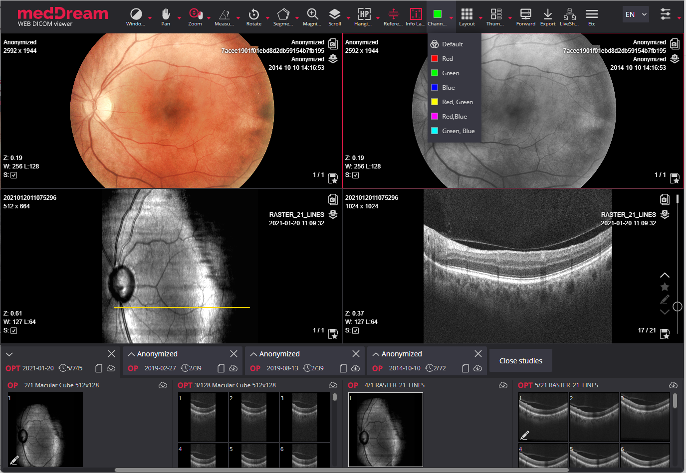
The MedDream DICOM Viewer proposes the possibility to view ophthalmic images from ophthalmology diagnostic and surgical devices such as retinal/fundus cameras, etc. allowing the use of ophthalmology PACS server as a retinal imaging archive. The DICOM Viewer for retinal imaging includes a wide range of features, standard tools, measurements, and specific tools for ophthalmology: Color channels, Reference lines.
Color channels tool to digitally apply monochromatic filters for the primary colors as well as for the secondary colors to enhance the visual contrast of anatomical details. This tool aims at highlighting a color component (red, green or blue) or a combination of them (red & green, red & blue, green & blue) by showing selected color in white shades and other colors in black.
Also DICOM Viewer supports most common modalities for ophthalmology:
Supported modalities for ophthalmology:
For more information about MedDream DICOM Viewer for retinal imaging download the Ophthalmology DICOM Viewer brochure.
The DICOM Viewer has wide range of specific measurements for measuring the distance, angle, Cobb angle, markers for spine labeling, which are important for orthopedic imaging. The specific orthopedic measurements are:
The DICOM Viewer supports the most common modalities for orthopedy: CT, CR, DX, MR, US, and other modalities and formats:
Supported modalities for orthopedy:

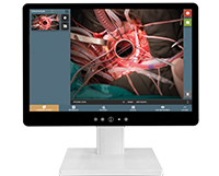
MedDream proposes the possibility to search, review and analyze medical videos from ophthalmology devices, microscopes, endoscopes, surgical video cameras, arthroscopes, echoscopes and other medical video sources. The Video module is integrated into the MedDream DICOM Viewer, allowing the use of PACS/VNA as a medical video archive.
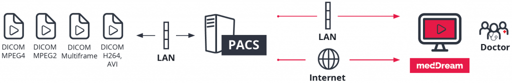
Video support:
Multi-frame support:
2023-12-15
Download the Linux zip package for: PacsOne, dcm4chee v2, dcm4chee v5, Conquest, Orthanc, DICOM Query/Retrieve, FileSystem, ClearCanvas, Google Cloud Healthcare, DICOMweb (WADO).
MedDream DICOM Viewer 8.3.1 functionality is a part of the MedDream 8.3.1 product.
Documentation
User Manual: General User Manual: Viewing functions Installation & Service Manual Integration API DICOM Conformance Statement SpecificationMaterials
Brochure Leaflet: Main Features Video Tutorials Certifications CE Declaration of Conformity Labeling on the device2023-12-15
Download the Windows setup.exe install file for: PacsOne, dcm4chee v2, dcm4chee v5, Conquest, Orthanc, DICOM Query/Retrieve, FileSystem, ClearCanvas, Google Cloud Healthcare, DICOMweb (WADO).
Quick Start Guides:
2023-12-15
Latest release of MedDream DICOM Viewer on Docker integrated into Orthanc DICOM server.
2023-12-15
Latest release of MedDream DICOM Viewer on Docker integrated into dcm4chee5 DICOM archive.
2023-12-15
Latest release of MedDream DICOM Viewer on Docker integrated into Azure Health Data Service.
MPR/MIP/3D reconstructions: Oblique functionality is replaced by Oblique MIST functionality. Oblique functionality will no longer be supported.
MedDream (Linux and Windows installations) works with JAVA 17 and FFMPEG modules. MedDream service must have the dedicated user with relevant rights. Please check information provided in the Install Manual.
MedDream v8.3.1 will be maintained for 1 year period from the release date.
For installation support or remote installation, please contact us at: info@softneta.com.
© 2007. Softneta. All rights reserved.