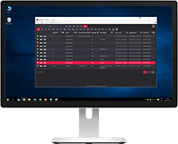
Software Vendors & OEMs
Ready to integrate web DICOM PACS Viewer and connectivity solutions

RETINAL IMAGING
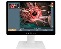
DICOM VIEWER
Ophthalmology DICOM Viewer ensures a way to search, view, analyze ophthalmic digital images, and make a diagnosis. Vendor-neutral DICOM Viewer proposes the possibility to view images, signals or videos from ophthalmology diagnostic and surgical devices. DICOM Viewer includes a wide range of features and measurements including specific tools for retinal images.

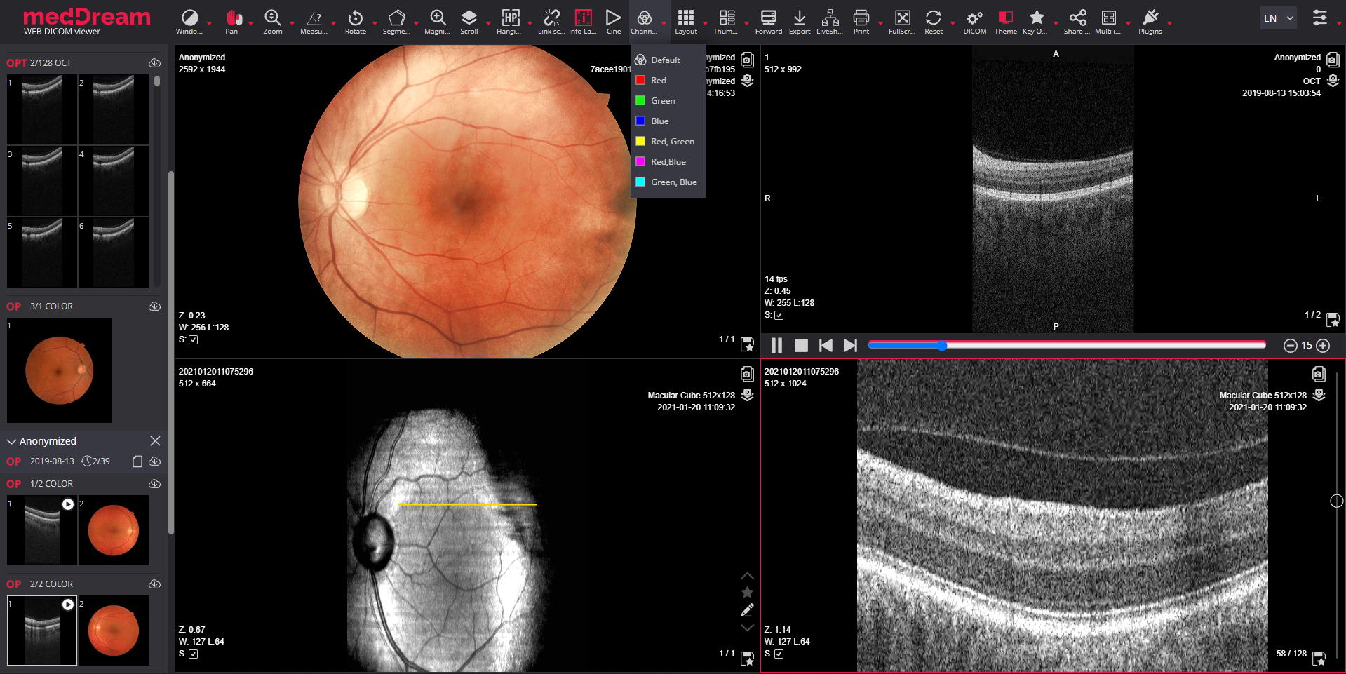


Retinal imaging with html5 MedDream DICOM Viewer ensures a way for ophthalmologists to search, view, analyze digital retinal images and make diagnosis from anywhere and on any device. This vendor neutral DICOM Viewer proposes the possibility to view ophthalmic images from ophthalmology diagnostic and surgical devices such as retinal/fundus cameras, etc. allowing the use of ophthalmology PACS server as a retinal imaging archive. DICOM Viewer supports modalities for ophthalmology: OCT, OPT, OP, OT, XC, SC, US and video formats. The DICOM Viewer for retinal imaging includes a wide range of features, standard tools and measurements but also specific tools for analyzing digital retinal images.
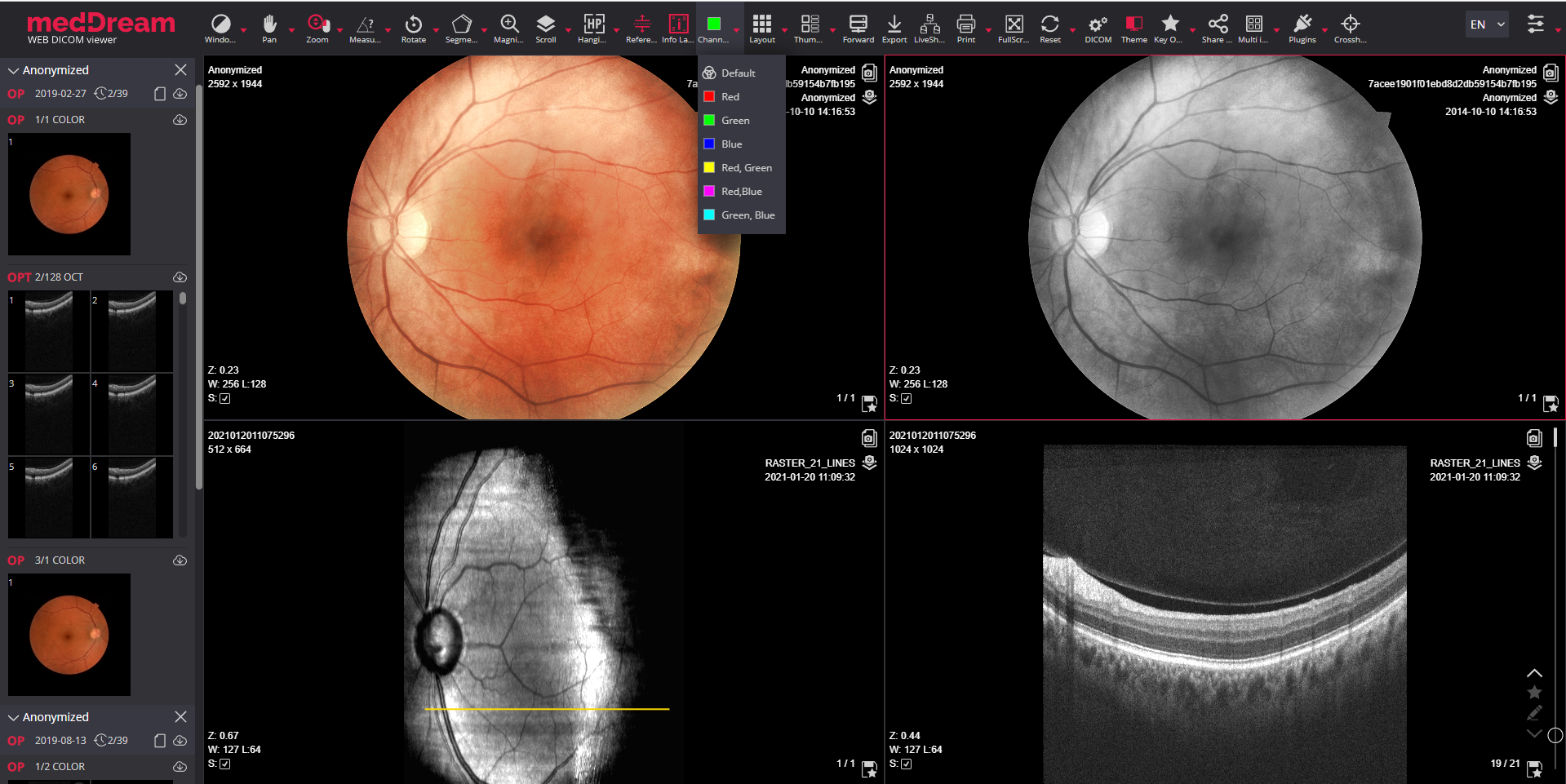
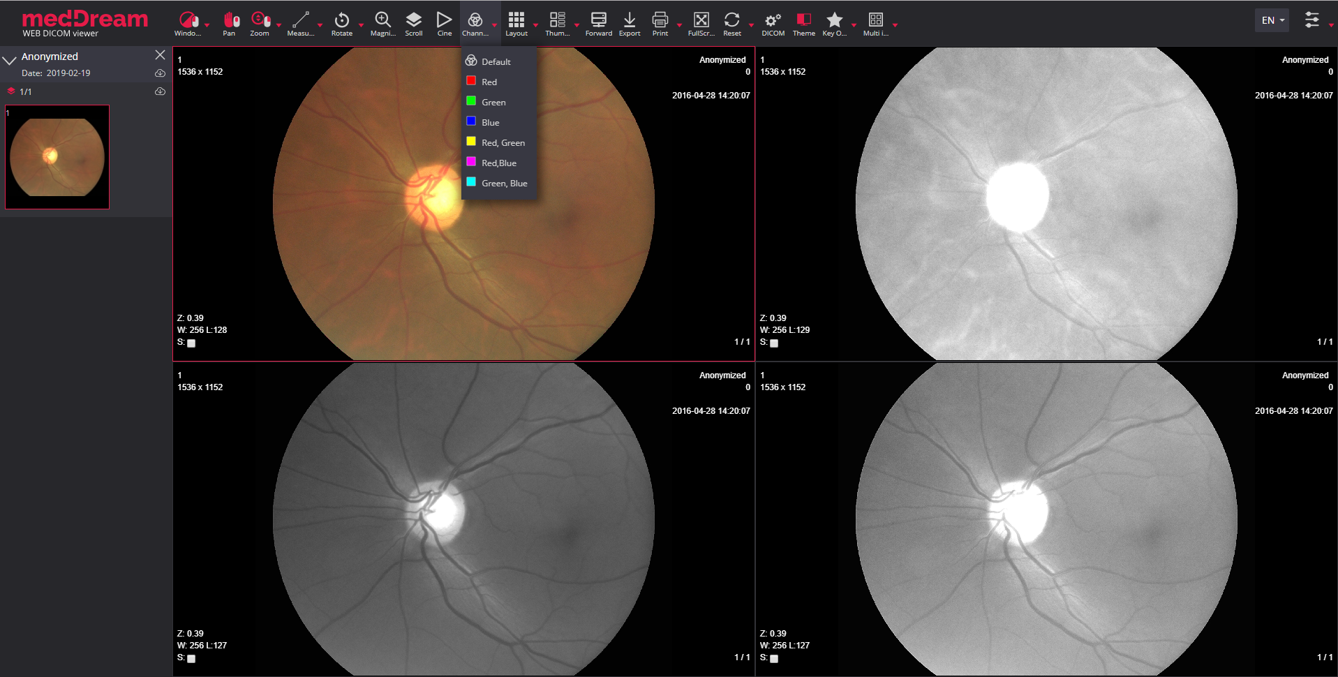
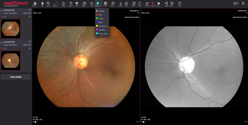
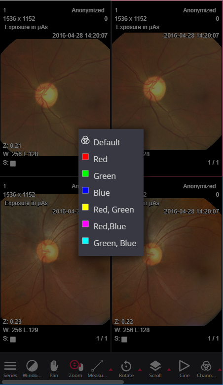
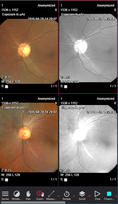
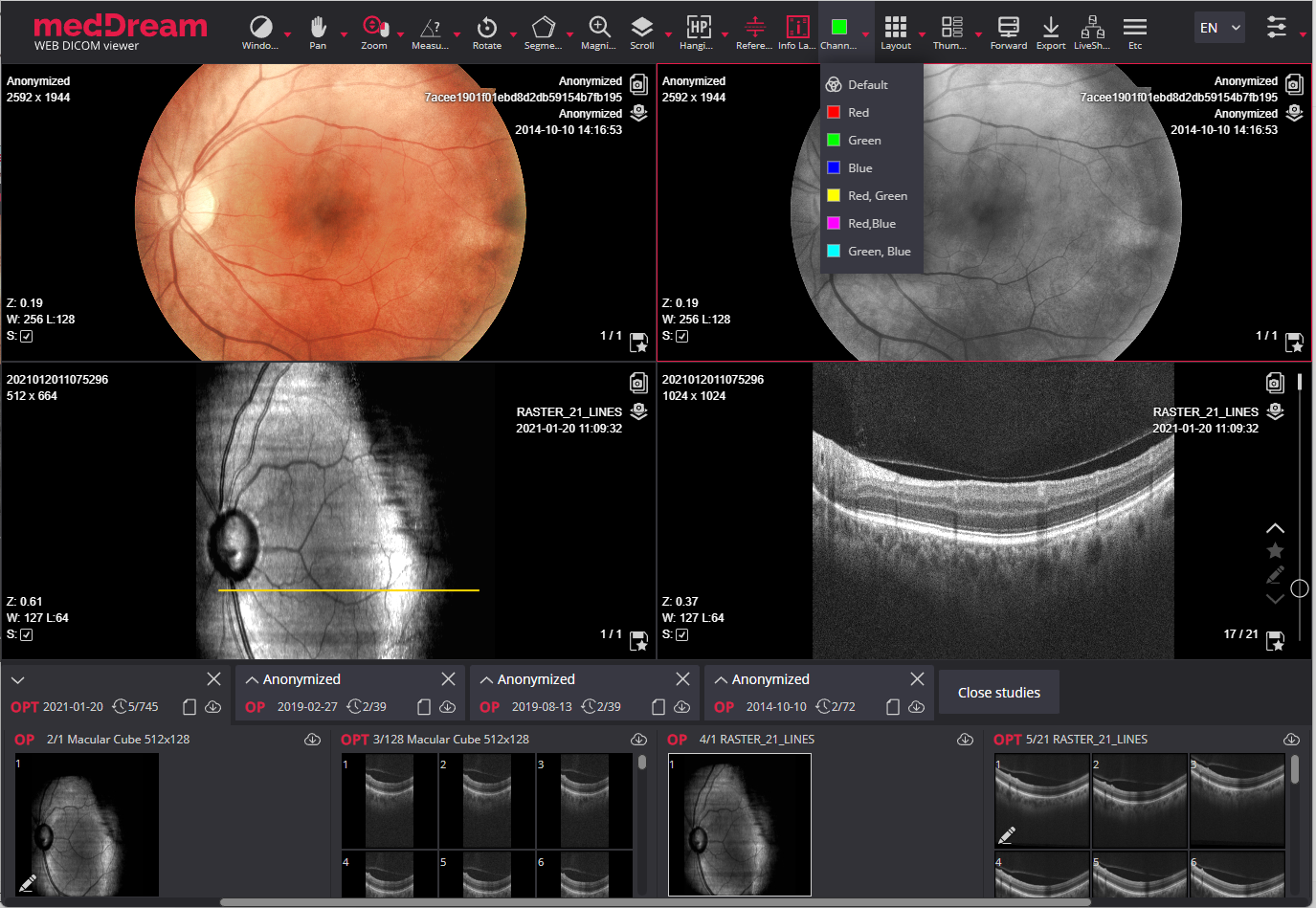
For retinal fundus imaging, ophthalmic photography or other visual spectrum images MedDream DICOM Viewer proposes a specific tools. Color channels tool for digital retinal imaging aims at highlighting a color component (red, green or blue) or a combination of them (red & green, red & blue, green & blue) by showing selected color in white shades and other colors in black.

Color channels. Digitally applying the monochromatic filters for the primary colors as well as for the secondary colors to enhance the visual contrast of anatomical details.

Reference lines. Overlaying reference lines allow to indicate the location of an image slice on another image of an intersecting pane.
The MedDream Ophthalmology DICOM Viewer has an extensive radiology tool set, which includes standard tools:

Window;

Pan;

Zoom;

Scroll;

Rotate/Flip;

Magnifier.
The MedDream DICOM Viewer measurements for ophthalmology are:

Line;

Angle;

Cobb angle;

Polyline;

Area;

Ellipse;

Rectangle;

ROI;

Flexpoly;

Pencil;
Arrow;

Text;

Continuous measurement;

Intensity;

Show angles;

Calibration line.

Save annotation.

Delete annotation.
The ophthalmology web DICOM Viewer has an advanced layout features, main of them are:

Layout;

Thumbnails;

Full screen;

Multi image;

Patient history;

Key Objects storing;

Hanging protocols.

Previous Hanging Protocol.

Next Hanging Protocol.
The MedDream Ophthalmology DICOM Viewer also has specific features:

SR support for SR documents;

PDF support for PDF files;

Non-dicom support for text, bmp, jpg, png, tiff, pdf, mp4 formats;

PR support for Presentation State annotations;
KO support for Key Objects selection and saving.
Also DICOM Viewer supports all modalities and video formats important for ophthalmology:
Supported modalities for diagnostic use:
Multi-frame support:
Video support:
Integration options:


For more information about MedDream DICOM Viewer for retinal imaging download the Ophthalmology DICOM Viewer brochure or read more about the MedDream WEB DICOM Viewer.
© 2007. Softneta. All rights reserved.